Elke Buschbeck
The visual system of predatory water beetle larvae (Thermonectus marmoratus)
The larvae of the sunburst diving beetle, Thermonectus marmoratus (Coleoptera: Dytiscidae) are highly efficient visually-guided predators. Their visual system consists of a cluster of six stemmata and one eye patch on each side of the head capsule (Fig.1). Histological investigations show that the organization of individual stemmata differs strongly from any eye that previously has been described. Based on general morphology, ultrastructural data, and the presence of actin-rich areas that are typical for rhabdoms we find that each eye is characterized by several retinas. The two most dorsal eyes on each side are relatively long and tubular (Fig. 2), and we have identified three spatially distinct areas with retinula cells in eye one. Because the organization of these tubular eyes is rather complex, we illustrate it with a 3D reconstruction (Fig. 3) The retinas are: (1) a band of two rows of rhabdoms along the medial side of the eye tube, (2) a flattened cone-shaped region towards the bottom of the tube which is formed by the rhabdoms of retinula cells that are oriented perpendicular to the light path, and (3) two horizontal rows of long rhabdoms parallel to the light path at the base of the tube. Particularly interesting is the second retina as its retinula cells are oriented 90° differently from retinula cells of other eyes. Based on preliminary behavioral experiments and optical measurements and calculations, we hypothesize that these receptors (in the most anterior eyes) are important for capturing prey and could allow the larvae to gauge the appropriate strike distance. The third retina is unique in that it only extends in one dimension. There are a few examples of extremely narrow retinas in other animals, all of which represent eyes that scan the environment. We currently investigate the possibility that diving beetle larvae use dorso-ventral head flexion to scan its environment, enlarging its visual field (movie). Another aspect in which we are interested is the evolutionary origin of these eyes. Fig. 4 illustrates the eyes of 5 different species of diving beetles.
This project has many possibilities for the involvement of students at all levels, and I am currently accepting new students into my lab. Any student interested in joining the lab should also look at the list of potential projects.
This project is in collaboration with Randy Morgan (Invertebrate Conservation Manager, Insectarium, Cincinnati Zoo and Botanical garden) and with Dr. John Layne of our department.
The following people are currently, or were recently working on this project:
- Karunyakanth Mandapaka
- Heather Hoy
- Sarah Sbita
- Elke Buschbeck
Figure 1
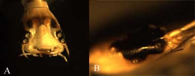
First instar larvae illustrating 6 lens eyes on each side of the head. (A) Frontal view, illustrating the two large pairs on each side. These are tubular and asymmetric. In the side view (B) the other 4, somewhat smaller eyes are visible as well.
Figure 2
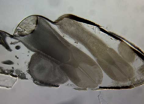
Saggital section of a first instar water beetle larvae illustrating the tubular shape of the two most anterior eyes.
Figure 3
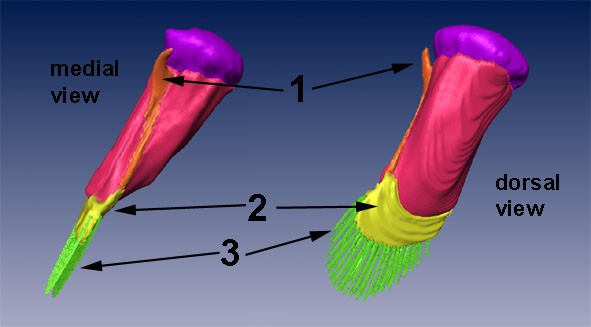
3D reconstruction of Eye 1 of the larvae of T. marmoratus. Three dedestinct retinas are visible (see numbers). Lens (purple), crystalline cone (magenta), lateral rhabdom (1;orange),rhabdomeric aggregation (2;yellow), and vertical retina (3;green)
Figure 4
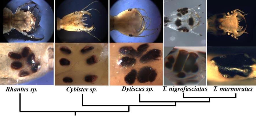
Head shape and eye placements in five dytiscid species that we currently keep in captivity and have been able to breed.